|
 |
 |


CASE 86: Leg-Located Rhabdomyosarcoma
- Hits: 161
A patient presented with swelling in the leg. Physical examination and radiological tests revealed a soft tissue mass, and a needle biopsy revealed a soft tissue sarcoma. The patient was diagnosed with tibial carcinoma (TCC). Scans revealed no metastases. Following radiotherapy, extensive resection of the patient's attached bone and existing knee prosthesis was performed, with a portion of the removed bone recovered using liquid nitrogen. Reconstruction was then carried out using a proximal tibia tumor resection prosthesis and a gastrocnemius muscle flap.
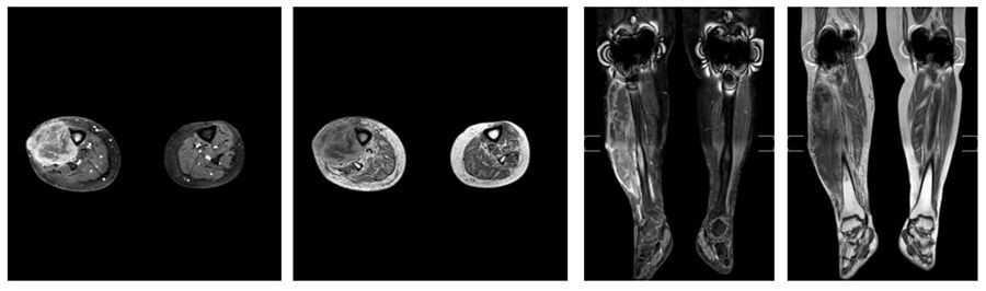
Preoperative: MRI shows an irregularly bordered, heterogeneous tumor tissue located anterolaterally on the crus, adjacent to the tibia, and causing a reaction in the medulla.
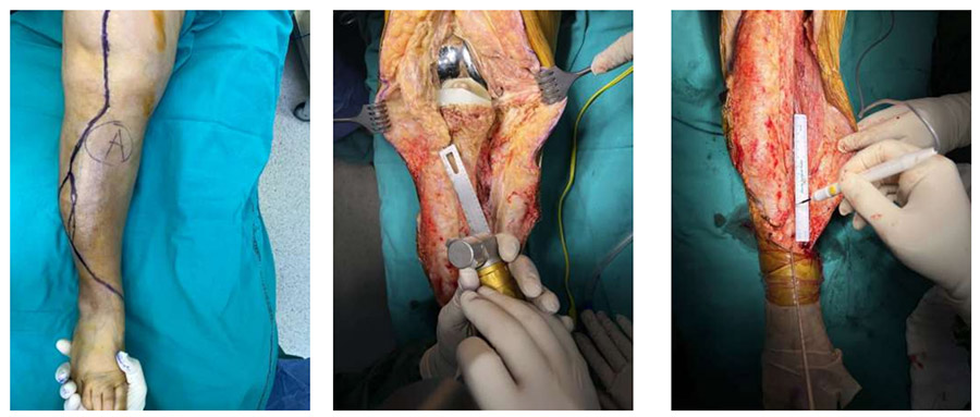
During the operation: Incision line to excise the biopsy tract, cutting the patellar tendon attachment site, determining the distal resection margin.
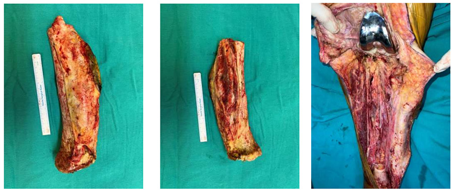
Surgical Procedure: The image shows the tumor tissue removed along with the skin and the bone it invaded, and the resulting cavity.
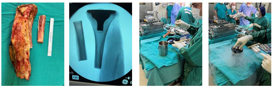
During the operation: Clinical and fluoroscopy images of the recovered bone and the application of liquid nitrogen are shown.
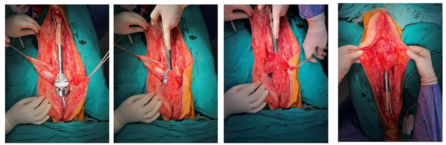
During the operation: The image shows the placement of a proximal tibia tumor resection prosthesis over the recovered bone, patellar tendon suturing, and closure with a gastrocnemius flap.
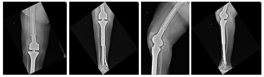
Postoperative: The X-ray shows a proximal tibia tumor prosthesis implanted after resection and subsequent recovery.