CASE 18: Hemipelvic Chondrosarcoma
- Hits: 41
- 27-year-old male patient
- The patient presented with complaints of left hip and groin pain accompanied by numbness in the foot and difficulty walking.
- The patient was diagnosed with chondrosarcoma via closed needle biopsy, and no metastases were detected in subsequent screenings.
- The patient underwent hip transposition with proximal femur tumor prosthesis following internal hemipelvectomy.
Preoperative: X-ray and 3CT scans show an obturator foromani originating from the left acetabulum and pubic arm, and irregularly bordered sclerotic tumor tissue filling the hemipelvis.

Preoperative: MRI shows tumor tissue in the same location, accompanied by a large, heterogeneous soft tissue component.

Preoperative: CT scan shows an irregularly bordered mass containing calcification causing bone surface damage.

During Surgery: Exposure and preservation of the hair follicles after sacral laminectomy.

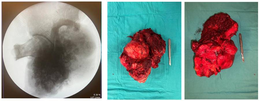
During the operation: Fluoroscopy and clinical view of the removed tumor tissue.
Post-operative: Healed incision lines are visible.

Postoperative: The X-ray shows hip transposition with proximal femur tumor prosthesis following internal hemipelvectomy.

