CASE 30: Chondrosarcoma located in the hip (proximal femur)
- Hits: 35
Our young male patient underwent extensive resection and reconstruction with tumor prosthesis due to chondrosarcoma located on the proximal femur.
Before the surgery: X-ray shows a lytic lesion, and MRI shows an irregularly bordered heterogeneous mass with surrounding edema.

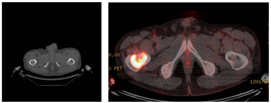
Before the surgery: PET-CT shows cartilage content and increased activity.
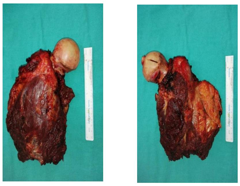
During the Surgery: Clinical image of the removed tumor tissue.
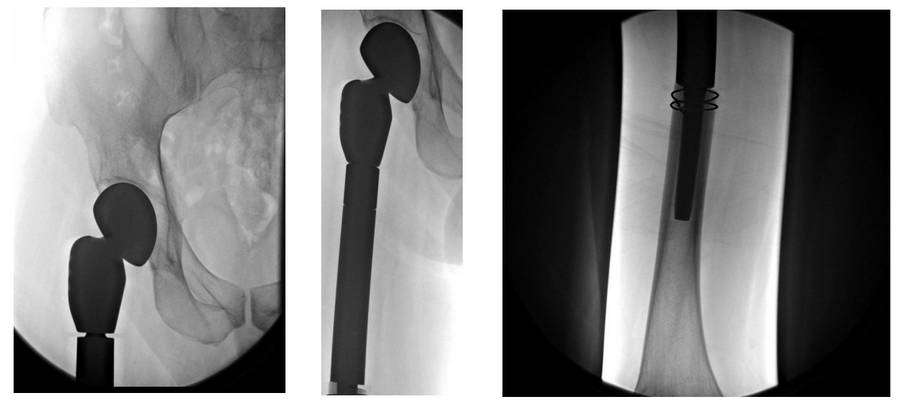
Post-Surgery: The X-ray shows the tumor prosthesis implanted after resection.