CASE 27: Chondrosarcoma located in the hip (femoral head)
- Hits: 37
Our young female patient underwent aggressive curettage, cementation, and fixation due to chondrosarcoma (Grade I) located in the femoral head.
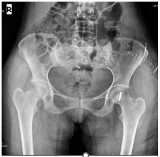
Preoperative examination: X-ray shows irregular borders, CT scan reveals thinning of the cortex and cartilage tissue, and MRI shows tumor boundaries.

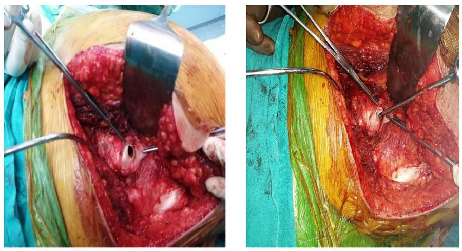
During Surgery: The image shows the tumor being removed, the resulting cavity being filled with bone cement, and then fixed with a headless screw.
Post-Surgery: The X-ray shows that after tumor removal, the resulting cavity was filled with bone cement and fixed with a headless screw.