CASE 14: Chordoma located in the sacrum
- Hits: 48
- 72-year-old female patient
- The patient presented with complaints of tailbone pain and constipation that had been present for a year.
- The patient who underwent closed needle biopsy for chordoma had no metastases detected in subsequent scans.
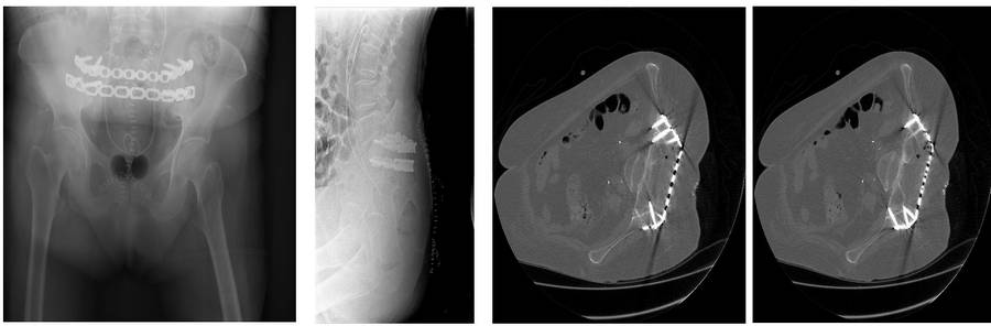
- After the tumor was removed with clean margins, the area was draped with prolene mesh to prevent hernia, and fixation was performed with two titanium plates.
Before the surgery: CT scans and MRIs show bone destruction in the sacrum and tumor tissue extending to the rectum.

During the Surgery: The image shows the rectum and sacral roots, the applied mesh, and the titanium plate after the tumor was removed in one piece.

During the Surgery: Clinical and fluoroscopy images of the removed tumor.

Post-surgery: The titanium plate that was implanted is visible in the X-ray and CT scan.