|
 |
 |


CASE 7: Humeral Chondrosarcoma
- Hits: 170
Our young male patient underwent aggressive curettage and cementum application due to chondrosarcoma (Grade I) located in the humerus.
Before the surgery: The X-ray shows tumor tissue containing irregular calcifications in the poximal humerus.

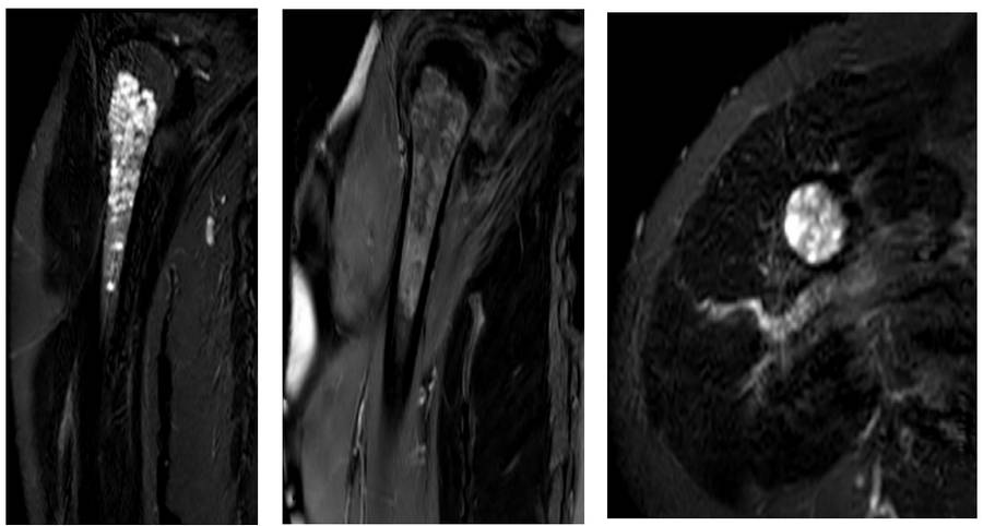
Before the surgery: The MRI shows a heterogeneous cartilage tumor filling the bone along the long segment.
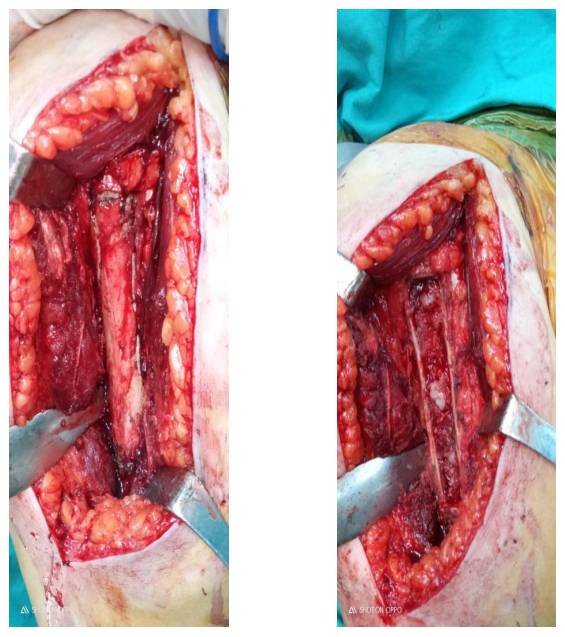
During the Surgery: The valve to be opened is marked, and after the valve is removed, the tumor tissue is seen.
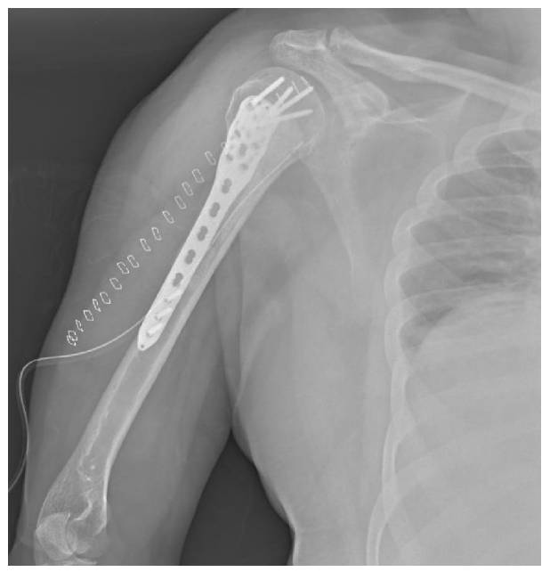
Post-Surgery: After the tumor is removed, the resulting cavity is filled with bone cement and reinforced with an anatomical plate.