|
 |
 |


CASE 34: Chondrosarcoma located in the shin (tibia)
- Hits: 134
A young female patient who underwent aggressive curettage, cementation, and fixation for tibia-located chondrosarcoma (Grade I).
Before the surgery: X-ray shows an irregularly bordered sclerotic mass on the lateral side of the proximal tibia.

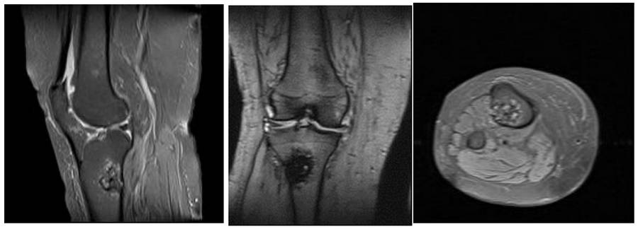
Before the surgery: MRI shows edema and cortical damage surrounding a heterogeneous mass.
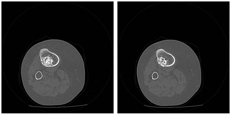
Before the surgery: The CT scan shows irregularities in the cartilage content and cortex.
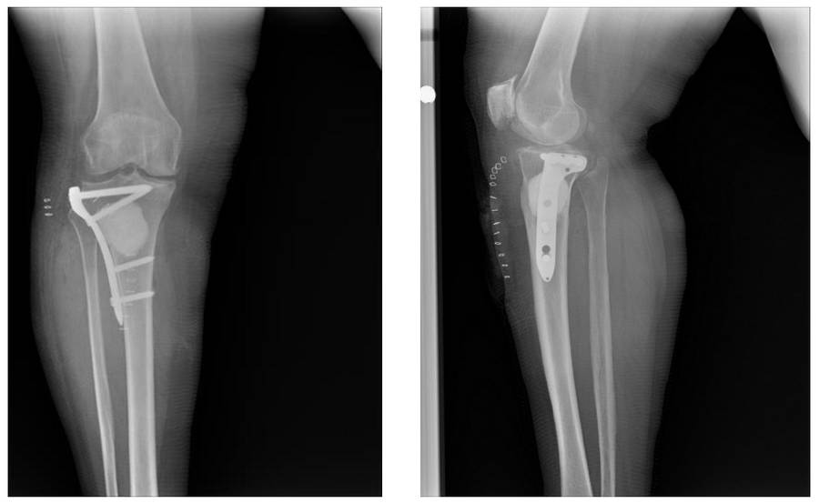
Post-Surgery: The X-ray shows fixation with bone cement and an anatomical plate filling the gap left by the removal of tumor tissue.