|
 |
 |


CASE 26: Proximal Femur Recurrent Chondrosarcoma
- Hits: 125
- 66-year-old female patient
- He underwent multiple surgeries due to proximal femoral chondrosarcoma.
- The prosthesis has been treated due to infection.
- The patient presented with complaints of swelling and pain in the left hip and thigh.
- The patient presented with a severe leg shortening and neurological deficit in the left leg.
- Internal hemipelvectomy with proximal femur resection was performed due to chondrosarcoma recurrence.
- A pre-prepared antibiotic spacer was placed in the resulting gap.
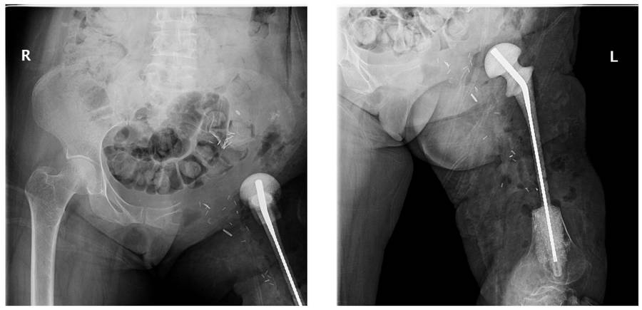
Before the surgery: The X-ray shows a spacer in place of the proximal femur absence, along with hip joint deformity and soft tissue calcification.

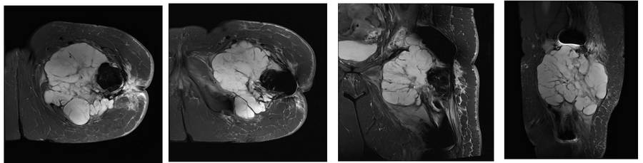
Before the surgery: The MRI shows a large tumor tissue with irregular borders, accompanied by a large soft tissue mass extending to the hip joint.
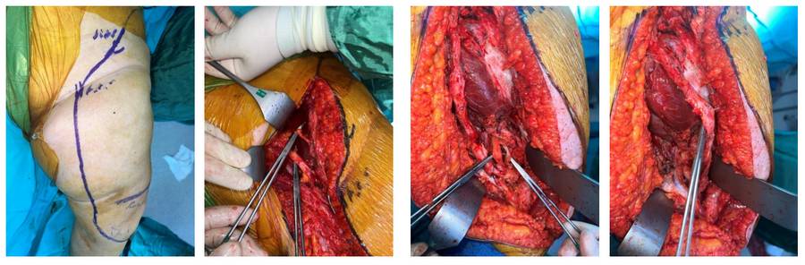
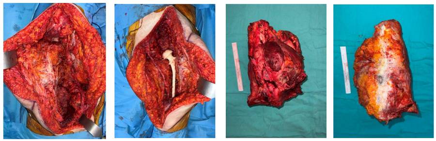
During the Surgery: The incision line shows the dissection and exposure of the vascular and nerve bundles.
Post-Surgery: The space created after resection, the application of an antibiotic spacer, and the clinical appearance of the removed tumor tissue.
Post-Surgery: The X-ray shows proximal femur resection and insertion of an antibiotic spacer after internal hemipelvectomy.