|
 |
 |


CASE 33: Chondrosarcoma located in the shin (tibia)
- Hits: 196
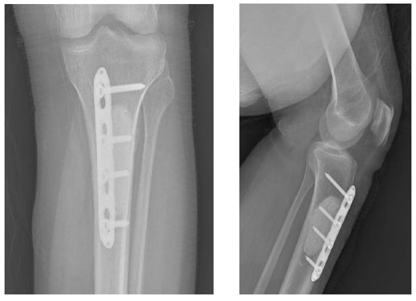
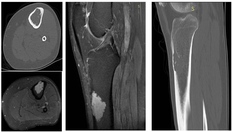
Our young male patient underwent aggressive curettage, cementation, and fixation due to tibia-located chondrosarcoma (Grade I).
Before the surgery: X-ray shows a lytic sclerotic mass in the proximal tibia.

Before the surgery: CT scan shows thinning of the cortex and cartilage tissue, while MRI shows irregular tumor borders.
Post-Surgery: The X-ray shows fixation with bone cement and plate applied to the site of the removed tumor.