|
 |
 |


CASE 32: Distal Femur Chondrosarcoma
- Hits: 260
Our adult female patient underwent aggressive curettage, cementation, and fixation for distal femoral chondrosarcoma (Grade I).
Before the surgery: The X-ray shows an irregularly bordered cartilage tumor located in the distal femur.

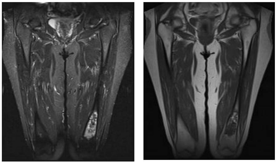
Before the surgery: The MRI shows a heterogeneous mass in the cortex lining the bone, causing thinning of the tissue.
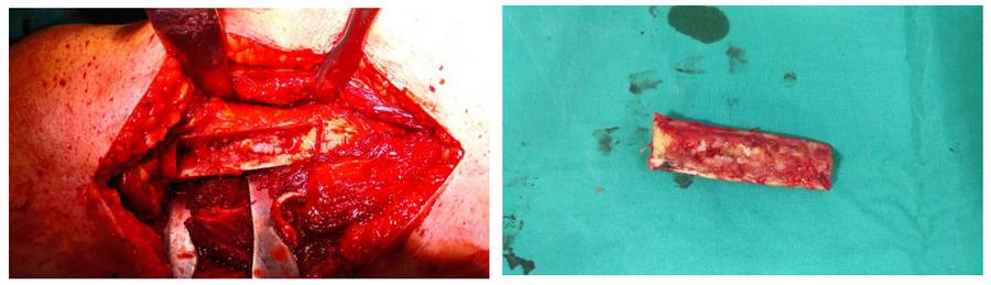
During the Surgery: The valve is open and tumor tissue is visible.
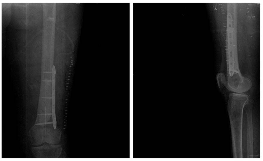
Post-Surgery: After tumor removal, the resulting cavity is filled with bone cement and reinforced with a plate.