|
 |
 |


CASE 82: Leg-Located Liposarcoma
- Hits: 181
A patient presented with complaints of swelling and pain in the leg. Physical examination and radiological tests revealed a soft tissue mass, and a needle biopsy revealed a soft tissue sarcoma. The diagnosis was made, and following radiotherapy, the tumor was removed along with the bone cortex with clean margins. The resulting cavity was filled with bone cement, and the surgery was performed to strengthen it with a titanium anatomical plate.
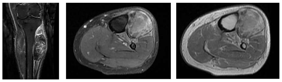
Preoperative: MRI shows edema surrounding an irregularly bordered, heterogeneous mass situated on the tibial cortex.
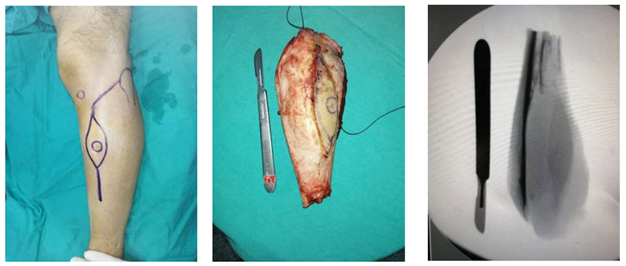
During the operation: The incision line to excise the biopsy tract, clinical and fluoroscopy images of the removed mass are shown.
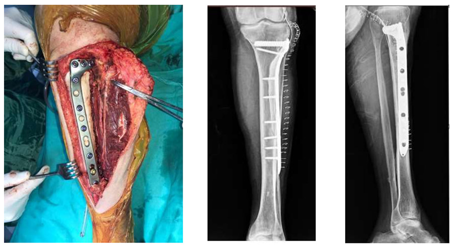
Surgery and Post-Surgery: The clinical and X-ray images show the reconstruction performed with cement and plate after the mass was removed.