|
 |
 |


CASE 1: Chondrosarcoma located in the shoulder blade (scapula)
- Hits: 185
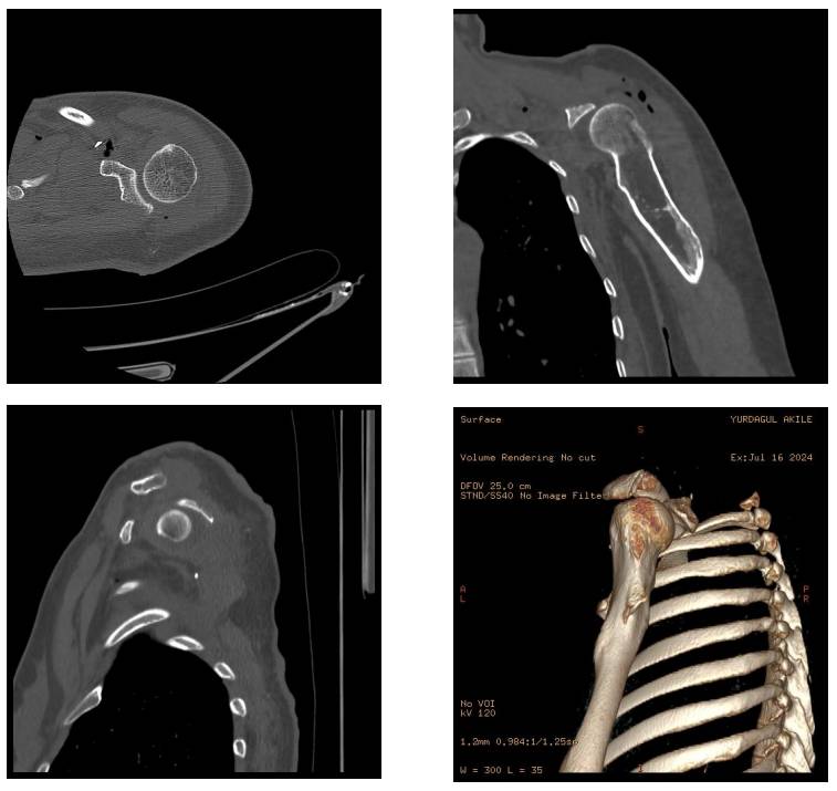
Our young female patient underwent scapulectomy due to chondrosarcoma developing in the scapula on the basis of multiple osteochondromatosis.
Before the surgery: The X-ray shows a giant, irregularly bordered mass of cartilage tissue originating from the scapula.

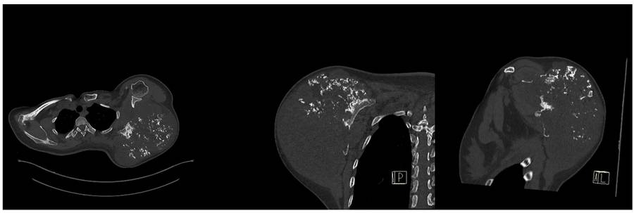
Before the surgery: The CT scan shows destruction of the scapula.
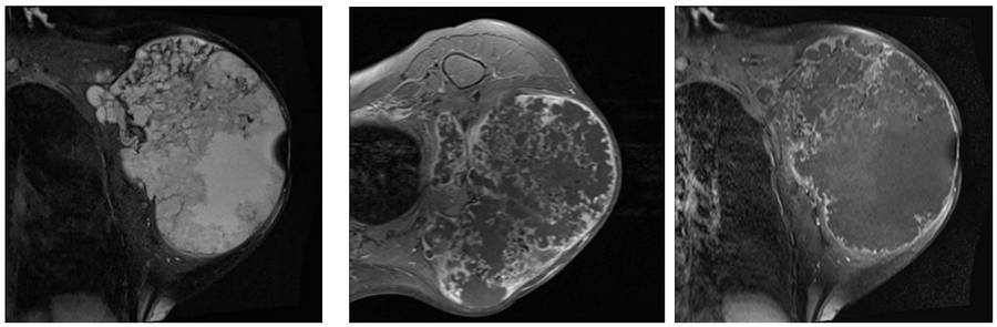
Before the surgery: The MRI shows the tumor to be heterogeneous and contain areas of necrosis.
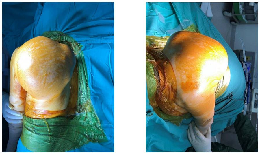
During the Surgery: Clinical appearance of the mass
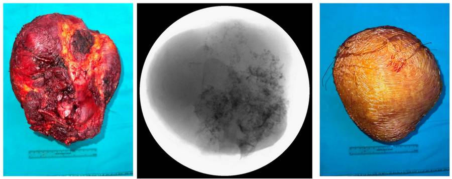
During the Surgery: Clinical and fluoroscopy images of the removed tumor tissue.
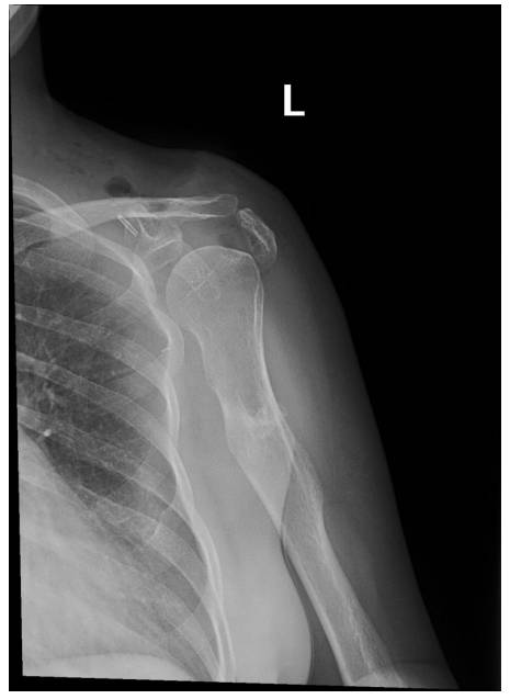
Post-Surgery: The X-ray shows that near-total scapulectomy was performed, excluding the glenoid joint surface.
Post-Surgery: The CT scan shows the resection margin and the remaining glenoid joint surface.