CASE 9: Osteosarcoma located in the lumbar, sacrum, and iliac wing
- Hits: 43
- 16-year-old female patient
- The patient, who had been experiencing pain and swelling for 10 months, underwent a biopsy abroad which revealed osteosarcoma, and received 4 rounds of chemotherapy.
- The patient, who had no metastases detected in the scans, underwent reconstruction with rod/screw+titanium cage+fibula after lumbo-sacro-pelvic resection.
- The patient, who experienced no complications during the surgery, was placed under close observation.
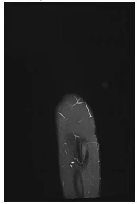
Before surgery: The MRI shows tumor tissue infiltrating the soft tissue, starting from the 4th lumbar vertebra and extending through the sacrum and iliac crest to the hip joint, causing bone destruction.

Before surgery: The MRI shows tumor tissue infiltrating the soft tissue, starting from the 4th lumbar vertebra and extending through the sacrum and iliac crest to the hip joint, causing bone destruction.

Before surgery: The MRI shows tumor tissue infiltrating the soft tissue, starting from the 4th lumbar vertebra and extending through the sacrum and iliac crest to the hip joint, causing bone destruction.
During the operation: Incision planning

During the operation: Clinical image of the removed tumor tissue.

During the operation: Reconstruction of the cavity created after tumor removal and fibula + titanium cage + rod/screw

Post-surgery: The X-ray shows the reconstruction procedure after resection.

