CASE 9: Sacroiliac Chondrosarcoma
- Hits: 40
- 46 year old man
- The pain and swelling in the lower back, which has been present for 5 months, has increased over time and makes it difficult to sit for long periods.
- A patient who underwent needle biopsy under CT scan was found to have chondrosarcoma, but no metastases were detected in subsequent scans.
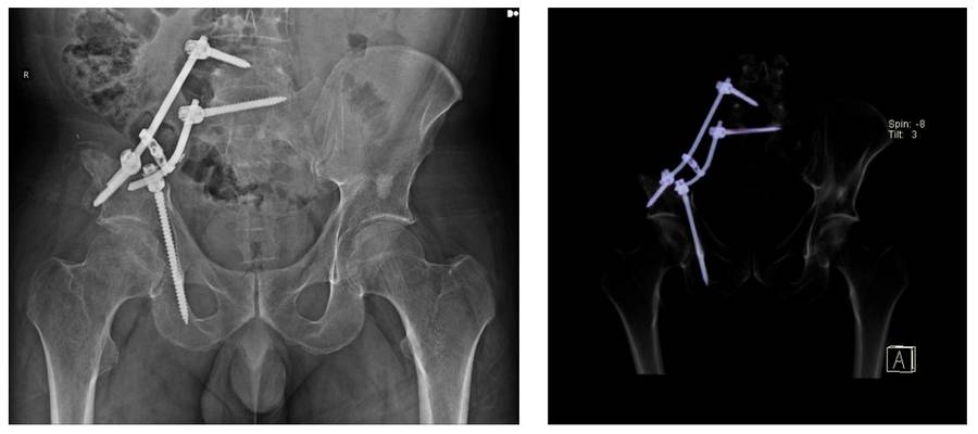
- After the tumor was removed with clean margins (sacroiliac resection), fixation was performed with rods and screws.
- The patient, who experienced no problems during or after the surgery, was placed under observation.
Preoperative scan: CT scan shows tumor tissue causing bone damage in the right iliac crest and sacrum, as well as a biopsy needle.

Preoperative: MRI shows tumor tissue with irregular borders and extension into soft tissue in the same location.

During surgery: Planning the incision line.

During the operation: Clinical and radiological images of the removed tumor tissue.

During the operation: Preservation of the lumbar and sacral roots and reconstruction after tumor removal.

Postoperative: The image shows fixation with rods and screws after sacroiliac resection.