CASE 5: Sacroiliac Soft Tissue Sarcoma
- Hits: 36
- 77-year-old male patient
- The patient presented with complaints of lower back/left leg pain and numbness in the left leg, which had been present for 6 months.
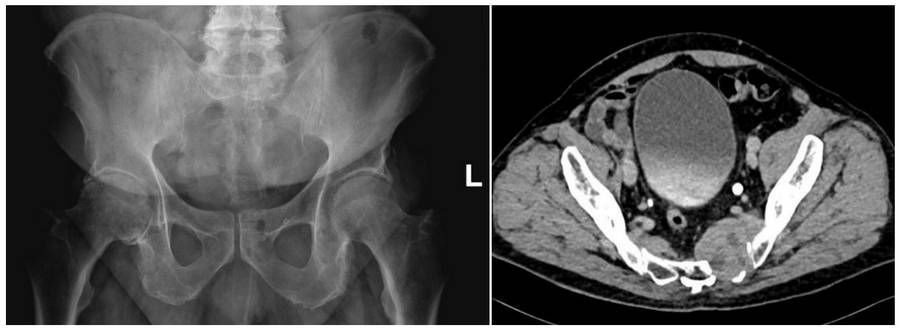
- Radiological examinations revealed a soft tissue mass originating from the left side of the sacrum, damaging the bone and extending to the hip.
- The patient, whose biopsy result showed a malignant peripheral nerve sheath tumor, showed no signs of metastasis in subsequent scans.
- After the tumor was removed with clean margins following radiotherapy (sacroiliac resection), the resulting space was reconstructed with lumbopelvic fixation.
Before the surgery: X-rays and CT scans show damage to the left side of the sacrum.
Before the surgery: The MRI shows tumor tissue originating from the left half of the sacrum and extending towards the hip.

During the operation: Fluoroscopy image of the removed tumor.

During the operation: Image of the removed tumor tissue and the reconstructed area.

Post-surgery: The X-ray shows fixation with a rod/screw after sacroiliac resection.

