|
 |
 |


CASE 83: Rhabdomyosarcoma located in the leg (cruris)
- Hits: 139
A patient presented with swelling in the leg, and physical examination and radiological tests revealed a soft tissue mass, which was then diagnosed as soft tissue sarcoma via needle biopsy.
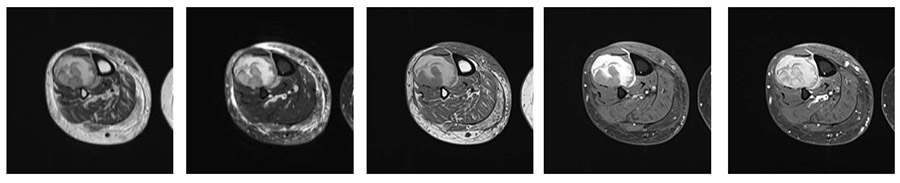
Preoperative: MRI shows an irregularly bordered, heterogeneous mass located in the midline of the crus, between the tibia and fibula.
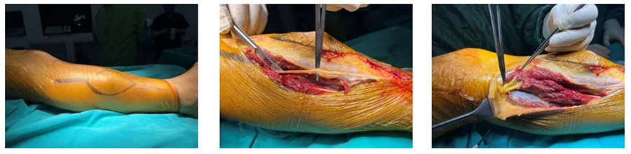
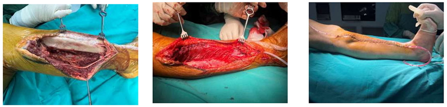
During the operation: The image shows planning the incision to excise the biopsy tract and exposing the sural and peroneal nerves.
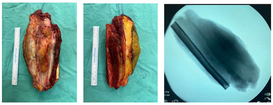
During the operation: Clinical and fluoroscopy images of the removed tumor tissue.
During the operation: After the tumor was removed, the resulting cavity is seen to be covered by muscle and skin.
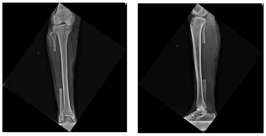
Post-operative: The X-ray shows that the middle portion of the fibula has been resected.