CASE 28: Chondrosarcoma located in the hip (proximal femur)
- Hits: 34
Our young male patient underwent wide resection due to chondrosarcoma located on the proximal femur on a background of multiple osteochondromatosis.
Before the surgery: X-rays show widespread osteochondromas around the hip and knee.

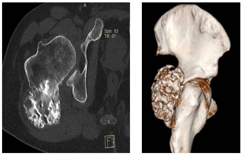
Before the surgery: The CT scan shows tumor tissue with irregular borders originating from the posterior proximal femur.
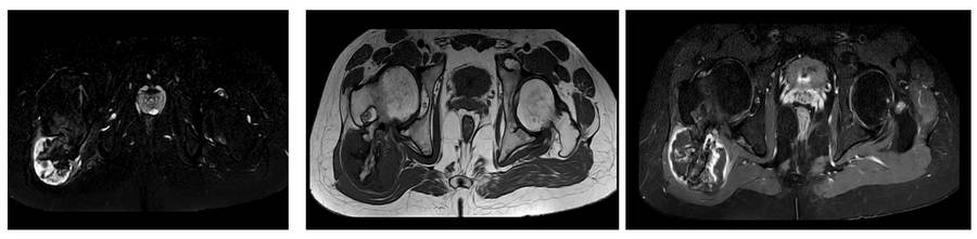
Before the surgery: The MRI shows a cartilage cap and edema around the tumor.
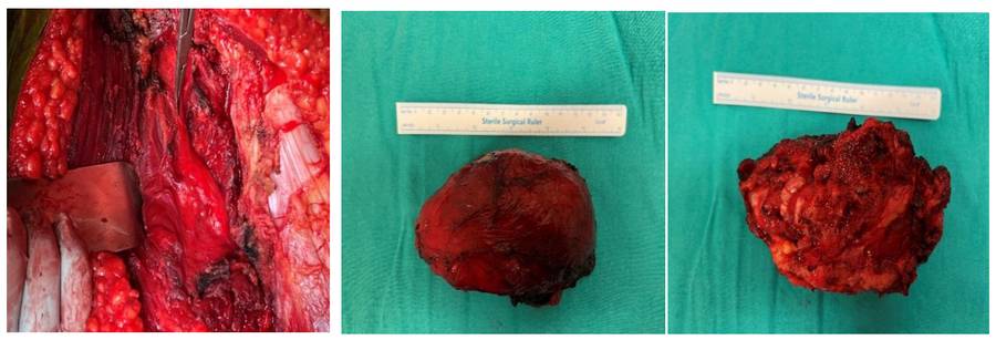
During the Surgery: The clinical image shows the sciatic nerve and the removed tumor tissue.
Postoperative: The X-ray shows that the tumor has been removed.

