CASE 40: A patient with colon cancer who underwent wide resection and tumor prosthesis due to the risk of fracture associated with metastasis to the right thigh (femur).
- Hits: 48
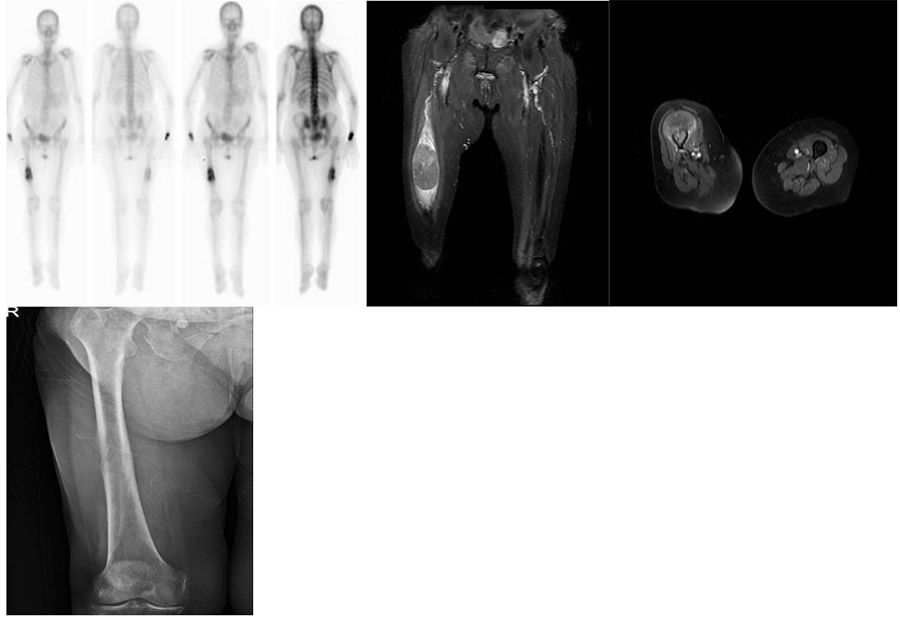
Before the surgery: X-ray shows bone destruction in the midline of the right femur, MRI shows an accompanying soft tissue component, and scintigraphy shows a single metastatic focus.
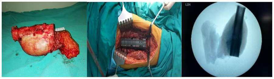
During the operation: The macroscopic and fluoroscopy images of the removed tumor are shown, along with the implanted tumor prosthesis.
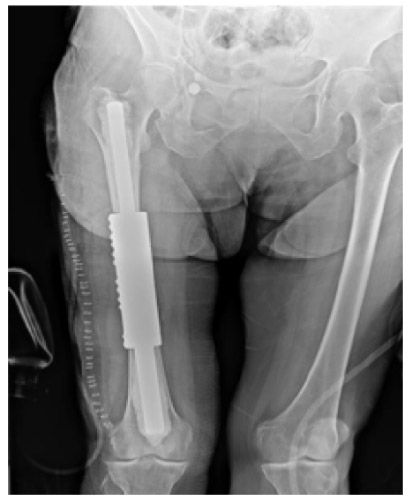
Post-surgery: The X-ray shows an intercalary cemented tumor prosthesis.
