CASE 36: Proximal Tibia Osteosarkom
- Hits: 41
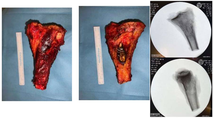
Our 70-year-old male patient underwent reconstruction with tumor prosthesis and gastrocnemius muscle flap after wide resection for proximal tibia osteosarcoma.
Before the surgery: The X-ray shows a mass located in the proximal tibia causing irregularly bordered bone destruction.

Before the surgery: The MRI shows tumor tissue extending beyond the bone in the same location, causing widespread peripheral edema.

During the Surgery: Clinical and fluoroscopy images of the removed tumor tissue.
During the Surgery: Tumor prosthesis, patellar tendon, gastrocnemius flap and closure.

Post-Surgery: The X-ray shows the tumor prosthesis implanted after resection.

