|
 |
 |


CASE 18: Hip (Femoral Neck) Osteosarcoma
- Hits: 215
Our young female patient underwent extensive resection and reconstruction with tumor prosthesis due to osteosarcoma located in the left femoral neck.
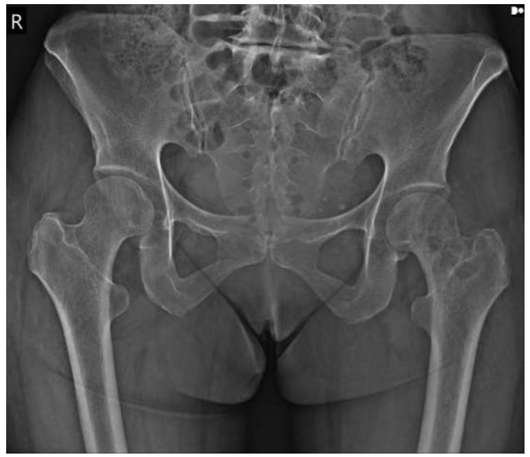
Before the surgery: X-ray shows an irregular lytic lesion located in the neck of the left femur.
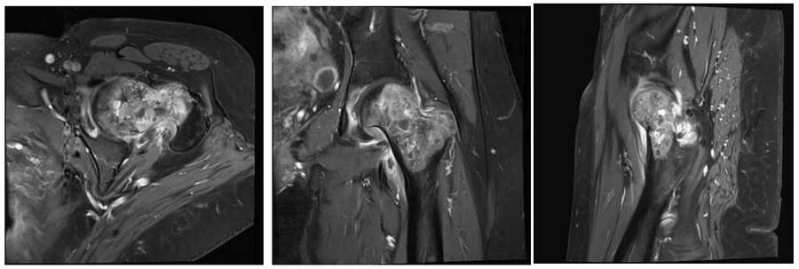
Before the surgery: The MRI shows irregular and dense tumor tissue filling the femoral neck, which enhances contrast and causes peripheral edema.
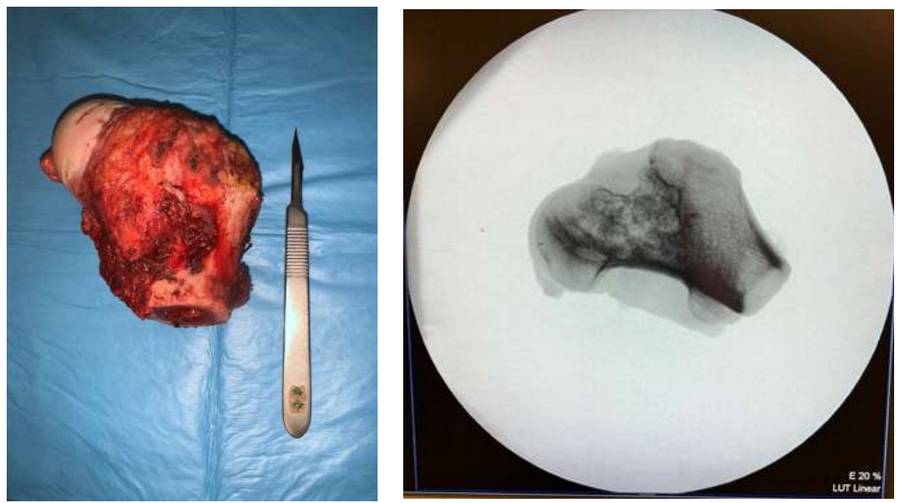
During the Surgery: Clinical and fluoroscopy images of the removed tumor tissue.
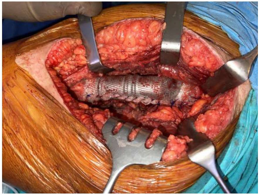
During Surgery: Suturing of the tumor prosthesis, applied after resection, to the surrounding soft tissues with prolene mesh.
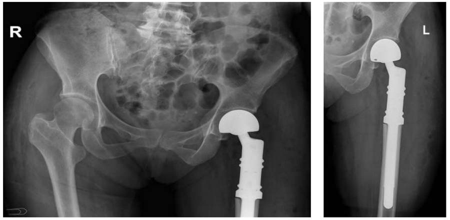
Post-Surgery: The X-ray shows the tumor prosthesis implanted after resection.