CASE 15: Osteosarcoma located in both hips (acetabulum)
- Hits: 37
- 19-year-old male patient
- He underwent 3 rounds of chemotherapy abroad for osteosarcoma located in the left acetabulum.
- No metastases were detected in the scans.
- The patient underwent extensive resection (Type II+III hemipelvectomy) followed by reconstruction (Lumic).
- At the 6th month of follow-up, metastases were detected in the right acetabulum and pubic arm of the patient. Since no additional metastases were detected in subsequent scans, the same surgical procedure was performed on the right side as well.
Before the surgery: MRI shows irregularly bordered tumor tissue extending into soft tissue and causing bone destruction in the left acetabulum and pubic arm.

During the Surgery: The image shows the cavity created after tumor removal, the removed tumor tissue, and the implanted prosthesis.

Post-Surgery: The X-ray shows sacroiliac fixation after resection (due to instability) and reconstruction with a Lumic prosthesis.

Before the surgery: The MRI shows tumor tissue that has damaged the bone and extended into the soft tissue in the right acetabulum and pubic arm.

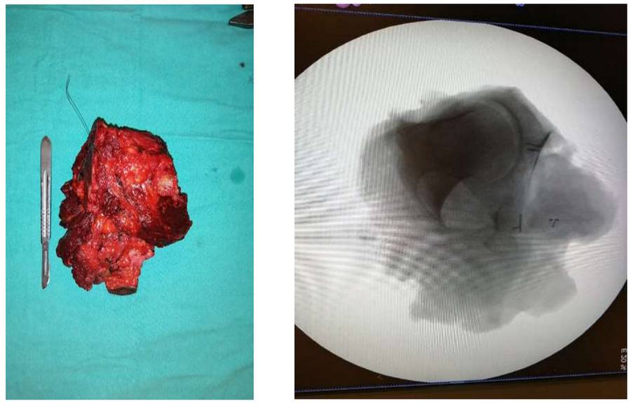
During the Surgery: The clinical and radiological images of the removed tumor tissue are shown.
Post-Surgery: The X-ray shows reconstruction with Lumic prosthesis after bilateral internal hemipelvectomy.

