|
 |
 |


CASE 14: Pelvic bone (iliac wing) osteosarcoma
- Hits: 294
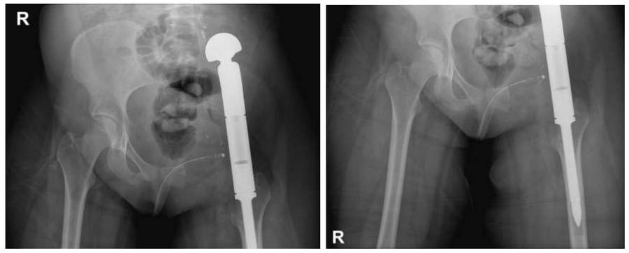
Our young female patient underwent extensive resection (internal hemipelvectomy) followed by reconstruction (hip transposition) with a modular tumor prosthesis due to osteosarcoma originating from the left pelvis and extending to the sacrum.
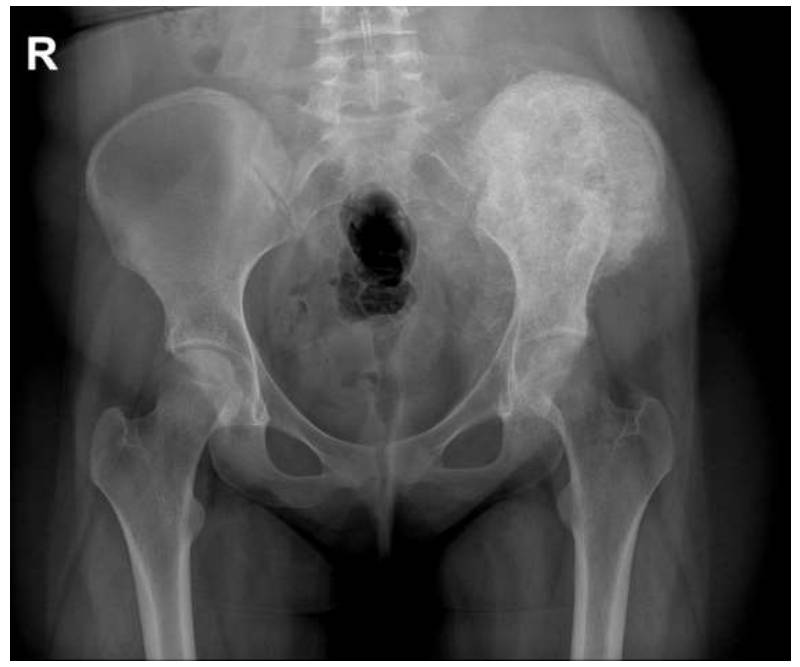
Before the surgery: The X-ray shows a sclerotic mass tissue completely covering the left iliac wing.
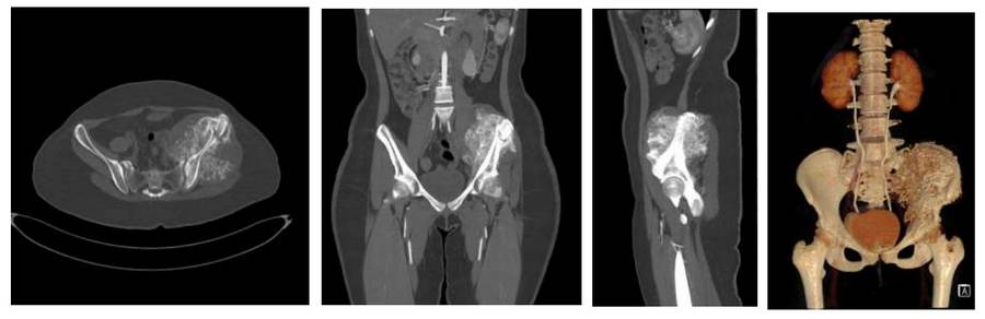
Before the surgery: CT scan shows irregular and well-defined tumor tissue originating from the left iliac wing.
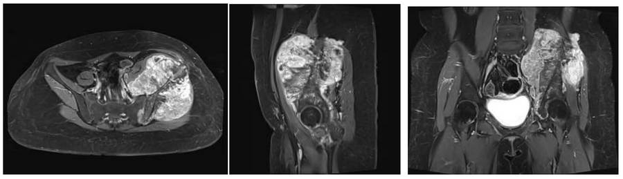
Before the surgery: The MRI shows tumor tissue in the same location, causing bone damage and extending into soft tissue.
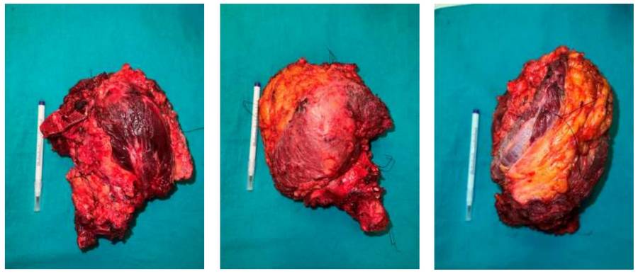
During Surgery: The clinical image of the removed tumor tissue is shown.
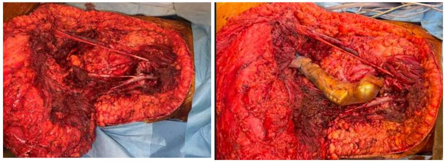
During the Surgery: The image shows the suturing of the resulting cavity and tumor prosthesis to the sacrum with prolene mesh after internal hemipelvectomy.
Post-Surgery: The X-ray shows hip transposition achieved with a tumor prosthesis after hemipelvectomy.