|
 |
 |


CASE 27: Osteosarcoma located on the distal femur
- Hits: 174
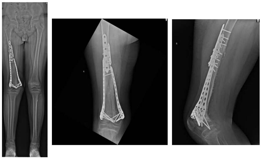
Our 11-year-old male patient underwent extensive resection followed by liquid nitrogen recovery and combined reconstruction with vascularized fibula due to distal femoral osteosarcoma.
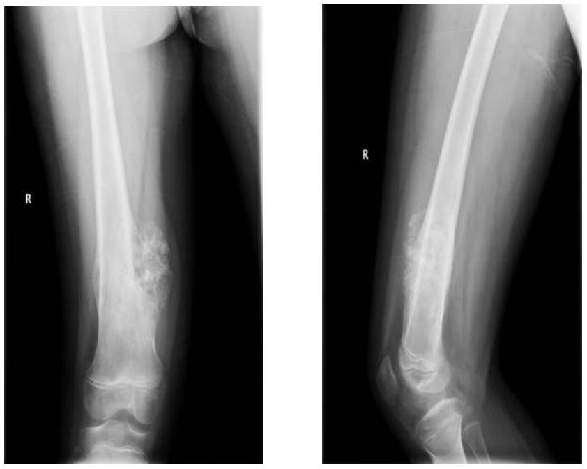
Before the surgery: The X-ray shows an irregularly bordered tumor mass causing bone destruction in the medial distal femur.
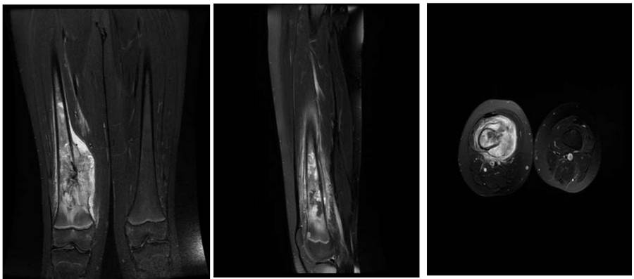
Before the surgery: The MRI shows tumor tissue located medially in the distal femur, causing bone destruction and extending into the soft tissue, accompanied by edema.
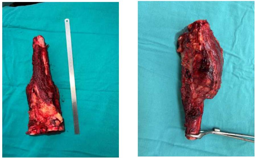
During the Surgery: Clinical image of the removed tumor tissue.
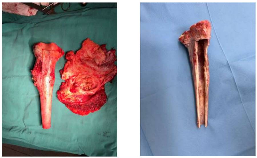
During the Surgery: Prior to bone recovery, the bone is separated from the surrounding soft tissue and medulla tumor tissue.
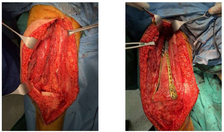
During the Surgery: Clinical images before and after reconstruction.
Post-Surgery: The X-ray shows the recovered bone tissue being fixed with a double plate proximally and distally after being combined with a vascularized fibula.