|
 |
 |


CASE 34: Osteosarcoma located in the knee (proximal tibia)
- Hits: 194
A 15-year-old male patient underwent extensive resection followed by reconstruction with tumor prosthesis and muscle flap due to osteosarcoma located in the proximal tibia.
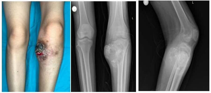
Before the surgery: Clinically and on X-ray, the tumor has an irregular, bordered, and heterogeneous appearance, causing significant damage to the medial proximal tibia and extending into the soft tissue.
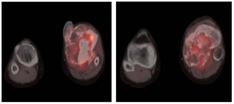
Before the surgery: PET-CT shows a lytic and sclerotic mass with heterogeneous and irregular borders and increased activity, causing significant damage.
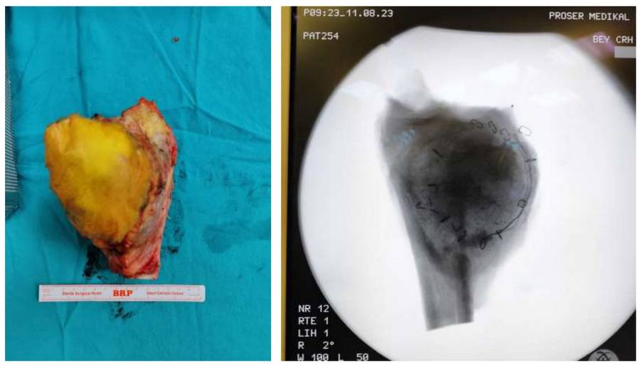
During the Surgery: Clinical and fluoroscopy images of the removed tumor tissue.
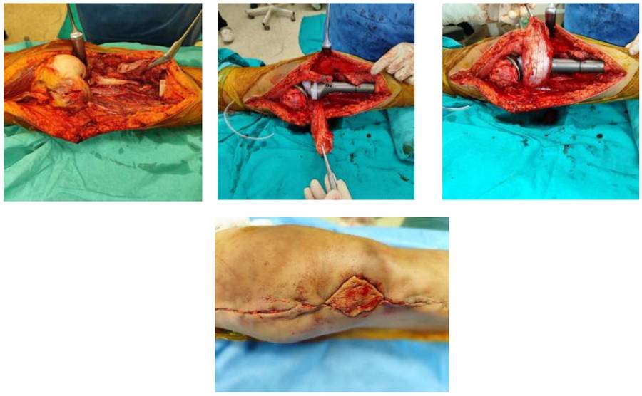
During the Surgery: The images show the procedure of filling the void created after tumor removal with a prosthesis, and applying a gastrocnemius muscle flap and skin graft after suturing the patellar tendon.
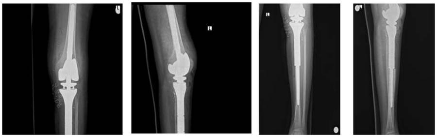
Post-Surgery: The X-ray shows reconstruction of the proximal tibia with a tumor prosthesis after resection.