|
 |
 |


CASE 19: Hip (Femoral neck) Osteosarcoma
- Hits: 167
A young adult male patient underwent extensive resection and reconstruction with a proximal femoral tumor resection prosthesis following chemotherapy for a pathological fracture due to osteosarcoma located in the femoral neck.
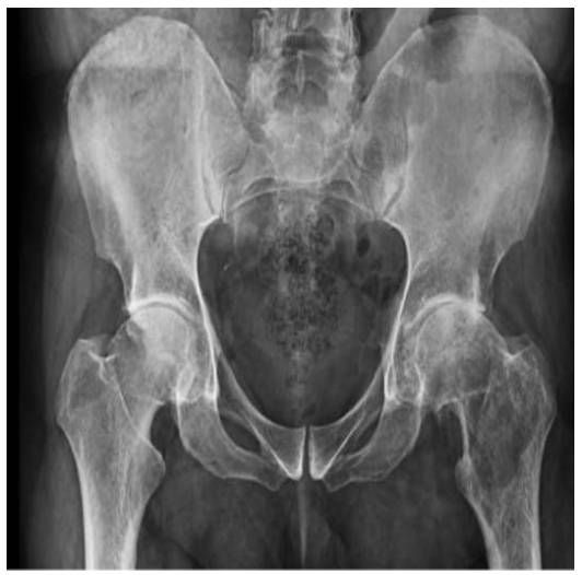
Before the surgery: X-ray shows a poorly defined lytic lesion in the femoral neck.
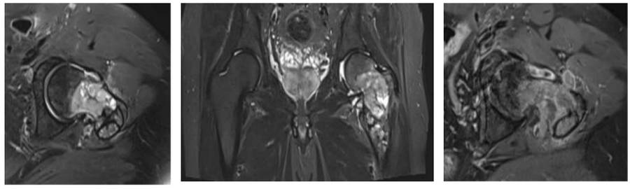
Before the surgery: The MRI shows tumor tissue located in the femoral neck, with irregular contents and extending beyond the bone.
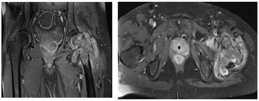
Before the surgery: MRI shows pathological fracture development and tumor spread to surrounding soft tissue.
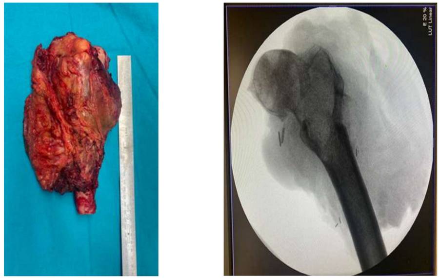
During the Surgery: Clinical and fluoroscopy images of the removed tumor mass.
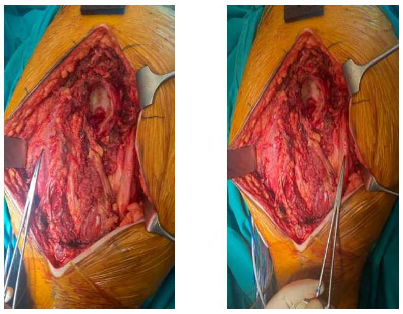
During the Surgery: After resection, the femoral artery, nerve and vein bundle are visible anteriorly, and the sciatic nerve is visible posteriorly.
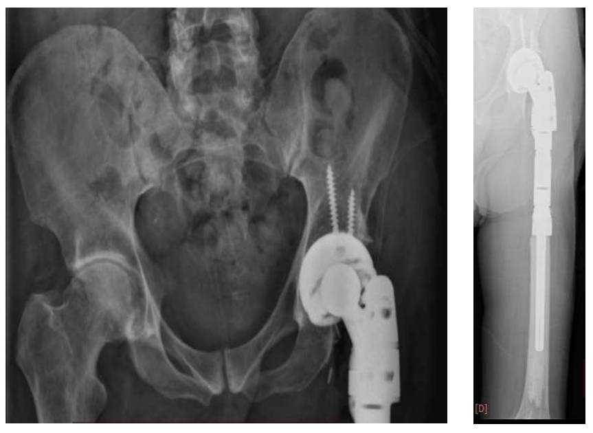
Post-Surgery: The image shows reconstruction using a proximal femur tumor prosthesis after resection.