CASE 32: Distal Femur Osteosarkom
- Hits: 41
Our female patient underwent extensive resection and reconstruction with tumor prosthesis due to distal femoral osteosarcoma.
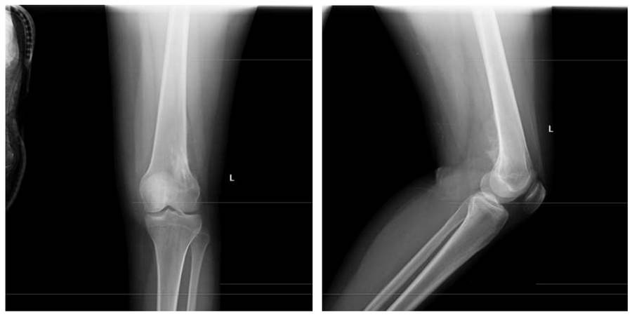
Before the surgeryThe X-ray shows an irregularly bordered mass located laterally in the distal femur, causing bone destruction and extending into the soft tissue.
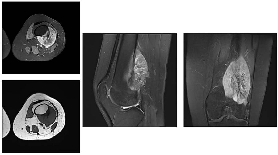
Before the surgery: The MRI shows tumor tissue adjacent to the vascular nerve bundle in the same location.
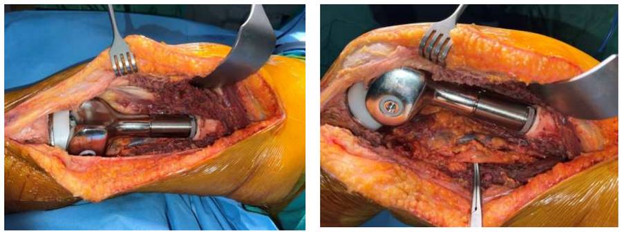
During the Surgery: Tumor prosthesis and vascular nerve bundle are visible.
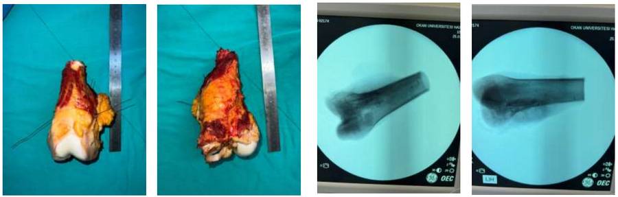
During the Surgery: Clinical and fluoroscopy images of the removed tumor tissue.
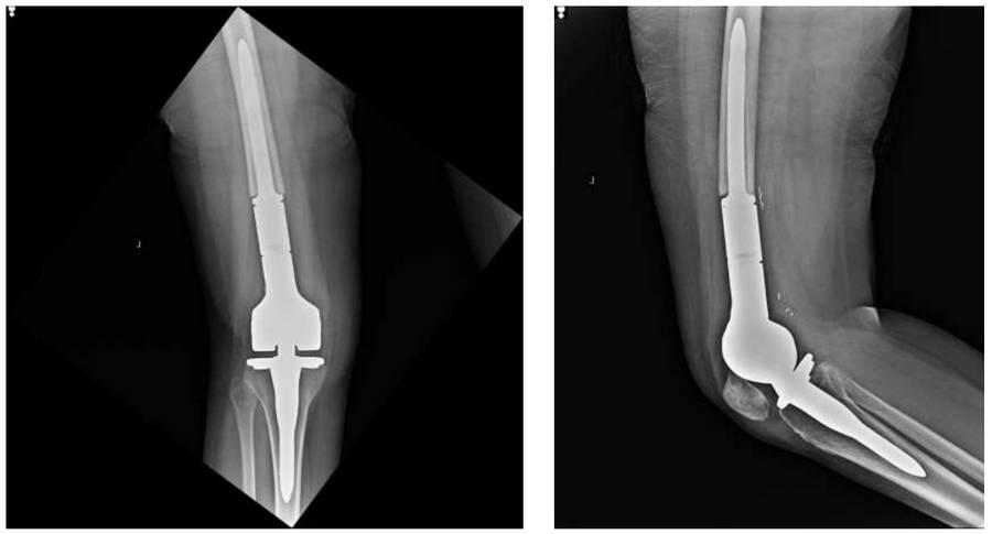
Post-Surgery: The X-ray shows the placement of a distal femur tumor prosthesis after resection.